Neuroanatomical atlas illustration plates from the 1786
'Traité d'Anatomie et de Physiologie' by Félix Vicq D'Azyr


[All the images above have been extensively background cleaned
and the colour saturation has been boosted to a modest degree]
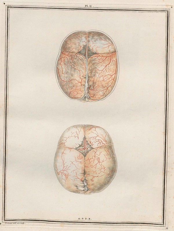
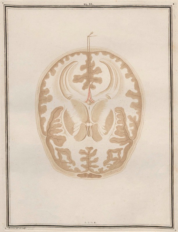
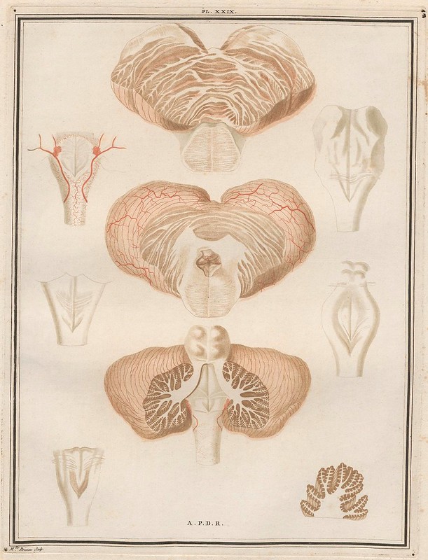
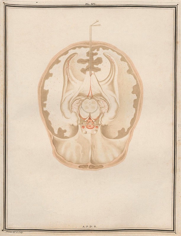
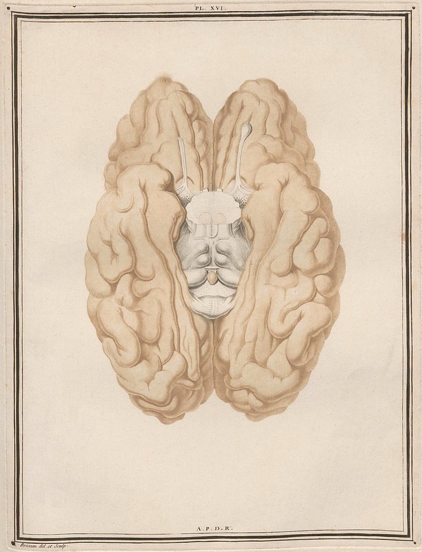
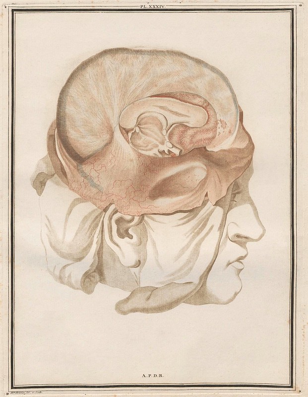
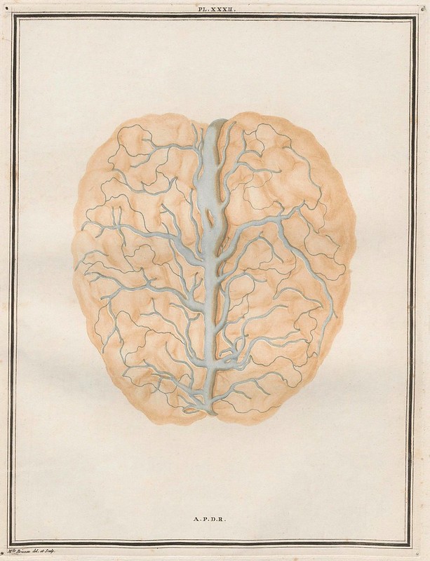
"Félix Vicq d’Azyr (1748-1794) was Queen Marie-Antoinette’s physician and a member of the French Academy of Sciences and of the Academy of Medicine. He wrote the section on pathological anatomy in 'L’Encyclopédie' by Denis Diderot and Jean le Rond d’Alembert, and in 1786 published 'Traité d’Anatomie et de Physiologie', which was lauded as one of the most realistic works of neuroanatomy. [It was described as "the most accurate neuroanatomical work produced before the advent of microscopic staining techniques": Garrison-Morton^ / "The work was issued in eight fascicles. The publication of the planned multi-volume work was interrupted by the French Revolution and the author’s premature death"*]
To illustrate his treatise, Vicq d’Azyr called upon the engraver Alexandre Briceau and his daughter Angélique, who used aquatint, an intaglio printmaking technique. A layer or successive layers of acid-resistant resin are baked onto a copper plate, which is then immersed in nitric acid to create different tones in the unprotected areas between resin particles, depending on acid concentration and exposure time. The plate is inked and prints are made. This complex technique achieves the delicacy of wash-paints, crayons, watercolors, and pastels. The etcher makes three plates, using aquatint (ochre), point engraving (red), and cutting wheel (black)." [source]
[Vicq d’Azyr] found that his dissections of the brain were facilitated by first hardening the brain in alcohol. He identified accurately for the first time many of the cerebral convolutions, along with various internal structures of the brain. He rediscovered the white line in calarine cortex and described the mammillothalmic tract which still bears his name, as well as the central sulcus with the pre- and postcentral convolutions and insula twenty years before Reil and Rolando." [source]
- 'Traité d'Anatomie et de Physiologie' by Félix Vicq d'Azyr was recently uploaded by Universitätsbibliothek Heidelberg (click 'Einband' and then 'Vorschau' for thumbnail pages)
- Félix Vicq d’Azyr at Bibliothec Systema Naturae : Hagströmer Medico-Historical Library. [W]
- [PDF] : Felix Vicq d'Azyr: Anatomy, Medicine and Revolution by André Parent IN: The Canadian Journal of Neurological Sciences.
- Medicographia: Body painting: five centuries of French anatomical illustrations.
- Long biography (in French) from a Parisian health university.
- Anatomia 1522-1867 : Anatomical Plates from the Thomas Fisher Rare Book Library (U Toronto).
- Historical Anatomies on the Web (National Library of Medicine).
- A copy of the book sold for >$20,000 a few years ago.
- Works by Vicq d'Azyr at Worldcat.
- Works by Vicq d'Azyr at the Internet Archive.
- Portraits of European Neuroscientists.
- Just because I found it: Bainbridge College biology course page with links to many different neuroanatomical atlas images.
- Previously: medicine :: science.
No comments:
Post a Comment